Home
/ Sketch And Label Of A Cross Section Of A Long Bone, 1 Schematic Drawing Of A Longitudinal Section Through A Long Bone Download Scientific Diagram - This is an online quiz called long bone anatomy.
Sketch And Label Of A Cross Section Of A Long Bone, 1 Schematic Drawing Of A Longitudinal Section Through A Long Bone Download Scientific Diagram - This is an online quiz called long bone anatomy.
Sketch And Label Of A Cross Section Of A Long Bone, 1 Schematic Drawing Of A Longitudinal Section Through A Long Bone Download Scientific Diagram - This is an online quiz called long bone anatomy.. Anatomy of a long bone 1. The end of a growing tibia, cut lengthwise*. Draw and label the following structures as they appear using the 10x objective o bone marrow o bony trabeculae activity 5.2.3: In these labeled examples, a human femur is represented without identifying many of the unique characteristics that help differentiate the femur bone from other bones in the human body. Create a drawing of the bone section in your laboratory journal and label the areas listed above.
There is a printable worksheet available for download here so you can take the quiz with pen and paper. Continue to label this drawing as you explore the inside of the bone. Looking at a bone in cross section, there are several distinct layered regions that make up a bone. Forms the larger rounded ends of long bones. Draw and label the following structures as they appear using the 10x objective o bone marrow o bony trabeculae activity 5.2.3:
Cartilage Bone Ossification The Histology Guide from www.histology.leeds.ac.uk Smartdraw includes 1000s of professional. Only the bottom portion of this bone extends as far as the hoof capsule. The cut line is called a cutting plane, and can be done in several ways. The diaphysis is the tubular shaft that runs between the proximal and distal ends of the bone. Bone test anatomy and physiology 12 photos of the bone test anatomy and physiology anatomy and physiology bone lab test, anatomy and physiology bone markings test, anatomy and physiology bone practical test, anatomy and physiology bone tissue test, anatomy and physiology test on bone tissue, bone, anatomy and. Forms the larger rounded ends of long bones. Create a drawing of the bone section in your laboratory journal and label the areas listed above. Create a drawing of the bone section in your laboratory journal and label the areas listed above.
There is a printable worksheet available for download here so you can take the quiz with pen and paper.
Anatomy of a long bone 1. Long bones have a thick outside layer of compact bone and an inner medullary cavity containing bone marrow. A section view is a view used on a drawing to show an area or hidden part of an object by cutting away or removing some of that object. Also known as the middle phalanx, the short pastern bone sits on top of the articulating joint of the pedal bone and underneath the long pastern bone. Terms in this set (3) epiphysis. Create a drawing of the bone section in your laboratory journal and label the areas listed above. Bone test anatomy and physiology 12 photos of the bone test anatomy and physiology anatomy and physiology bone lab test, anatomy and physiology bone markings test, anatomy and physiology bone practical test, anatomy and physiology bone tissue test, anatomy and physiology test on bone tissue, bone, anatomy and. In these labeled examples, a human femur is represented without identifying many of the unique characteristics that help differentiate the femur bone from other bones in the human body. The ends of a long bone contain spongy bone and an epiphyseal line. The diaphysis is the tubular shaft that runs between the proximal and distal ends of the bone. A long bone has a shaft and 2 ends. The diaphysis of a long bone is composed of bone tissue while the epiphysis is made of bone tissue. Cross section of a long bone.
End of a long bone. Lamellar bone makes up the compact or cortical bone in the skeleton, such as the long bones of the legs and arms. The diaphysis is the tubular shaft that runs between the proximal and distal ends of the bone. The following slides will help show the several methods or types of section views A typical long bone shows the gross anatomical characteristics of bone.
Long Bone Cross Section Diagram Quizlet from o.quizlet.com The diaphysis is the tubular shaft that runs between the proximal and distal ends of the bone. The little black spots are osteocytes. Osteons are oriented parallel to the diaphysis of the long bone. This is the long central shaft. In the space provided draw a longitudinal section of a long bone and label the following parte proximal epiphysis, distal epiphysis, diaphysis, metaphysis, medullary cavity, epiphyseal line 2. Sketch and label of a cross section of a long bone. Sketch and label of a cross section of a long bone. Diaphysis • shaft of the long bone.
Sketch and label of a cross section of a long bone.
It suggests that the bone will have equal strength in all directions. Area between the diaphysis and epiphysis at both ends of the bone. The little black spots are osteocytes. This is an online quiz called label the long bone. A long bone has two parts: External circumferential lamellae, osteon, central canal, perforating canals, lacuna, canaliculi, concentric lamellae. Marks should be deducted for shading or colouring. Cow and human long bones have a similar general structure. Osteons are oriented parallel to the diaphysis of the long bone. Lamellar bone makes up the compact or cortical bone in the skeleton, such as the long bones of the legs and arms. Continue to label this drawing as you explore the inside of the bone. Make a pencil sketch and use markers or colored pencils to add details. The cut line is called a cutting plane, and can be done in several ways.
Anatomy of a long bone 1. Use colored pencils to draw and label the following structures as they appear using the 40x objective, or by looking at an image from the internet. The cut line is called a cutting plane, and can be done in several ways. The diaphysis is the tubular shaft that runs between the proximal and distal ends of the bone. Cross section of a long bone.
Label The Parts Of A Long Bone from anatomycorner.com Osteons are oriented parallel to the diaphysis of the long bone. The diaphysis is the tubular shaft that runs between the proximal and distal ends of the bone. Cow and human long bones have a similar general structure. End of a long bone. Label lines should not cross ; Only the bottom portion of this bone extends as far as the hoof capsule. The ends of a long bone contain spongy bone and an epiphyseal line. Once we stop growing (between 18.
This is the long central shaft.
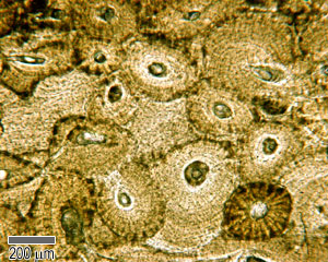
The diaphysis and the epiphysis. In the space provided draw a longitudinal section of a long bone and label the following parte proximal epiphysis, distal epiphysis, diaphysis, metaphysis, medullary cavity, epiphyseal line 2. This photo shows a cross section through bone. There is a printable worksheet available for download here so you can take the quiz with pen and paper. Label the haversian canal, osteocyte (mature bone cell) in lacuna, and canaliculi. Also known as the middle phalanx, the short pastern bone sits on top of the articulating joint of the pedal bone and underneath the long pastern bone. The ends of a long bone contain spongy bone and an epiphyseal line. Smartdraw includes 1000s of professional healthcare and anatomy chart templates that you can modify and make your own. The outside of a bone is covered in a thin layer of dense irregular connective tissue called the periosteum. Draw and label a longitudinal section of a long bone. The humerus is the long bone in the upper arm. Learners should accurately draw a long bone, resembling that in figure 6.24. The following slides will help show the several methods or types of section views

